1. From optical imaging to brain imaging
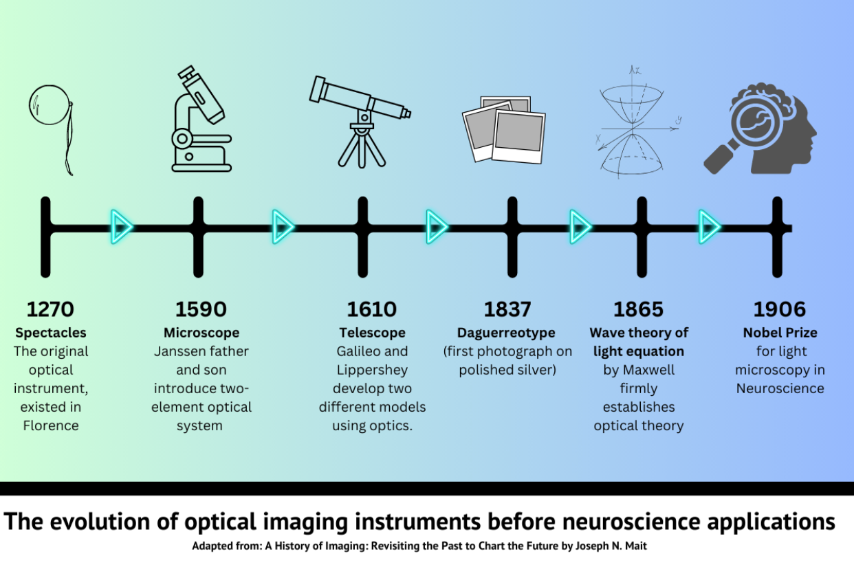
The use of light as an investigative tool for scientific research has been used by humankind for centuries. Optical instruments have been documented as far back as the 13th century, and today, “optics” form the very basis of many advanced research tools found in labs all over the world. Spectacles, which utilized a glass lens’s ability to magnify images, were the first practical optical instrument recorded (1). Over the next millennium, further developments included discoveries of glass types suitable for optical studies, photographs, the invention of the telescope and microscope, and ultimately a number of theoretical equations that have defined our understanding of how light can be used as a tool for research.
The scientific field of neuroscience has benefited hugely from the use of optics as an investigative tool. In relation to brain imaging, Camillo Golgi and Santiago Ramo´n y Cajal made a revolutionary discovery using light microscopy to trace neurons to structure the nervous system, ultimately winning the Nobel prize in 1906 (2). Investigations used today such as EEG, MRIs, PET scans, patch-clamp recordings and optical imaging all stem from this discovery made over 100 years ago.
2. Optical imaging in preclinical neuroscience
In preclinical neuroscience research, optical imaging offers an advantage compared to molecular biology techniques because it can offer the true physical features and functions of neurons in real-time (ref). Brain structure and functions studied in animal models allows for a better understanding of neural mechanisms such as cognition, sleep, emotion, mobility etc that cannot be easily studied in humans. These studies also help further our knowledge of nervous system diseases, and could potentially help unlock better treatments for humans affected by these diseases (3).
Optical imaging tools used in labs worldwide such as optogenetics, fiber photometry and fluorescence imaging all use a combination of light/lenses and genetic modifications or manipulations to study neuronal populations in the central nervous system. These techniques have contributed to a huge number of scientific discoveries in neuroscience, and as these technologies are evolving, more discoveries about the nervous system are being made annually.

3. Advancements & limitations in optical imaging for pre-clinical neuroscience
While the potential power of optical imaging had been theorised for decades, the technology and genetic modifications needed to carry out many neuroscience experiments has only recently been developed. Fluorescence imaging using calcium indicators has become an extremely powerful tool in recent years.. Fluorescence imaging miniscopes using advanced optical lenses (known as GRIN lenses) are allowing for more developed studies looking at the functions of individual single neurons, instead of just large populations of neurons (as seen in fiber photometry studies).
Some limitations faced by fluorescence imaging miniscopes have been:
- Developing thorough techniques to effectively image the activity of single neurons in real-time
- Developing functioning systems suitable for in vivo freely-moving studies
- Unable to access deep-brain regions due to damage to brain tissue caused by the implant (3)
4. Into the Future: Our Solution – Mightex
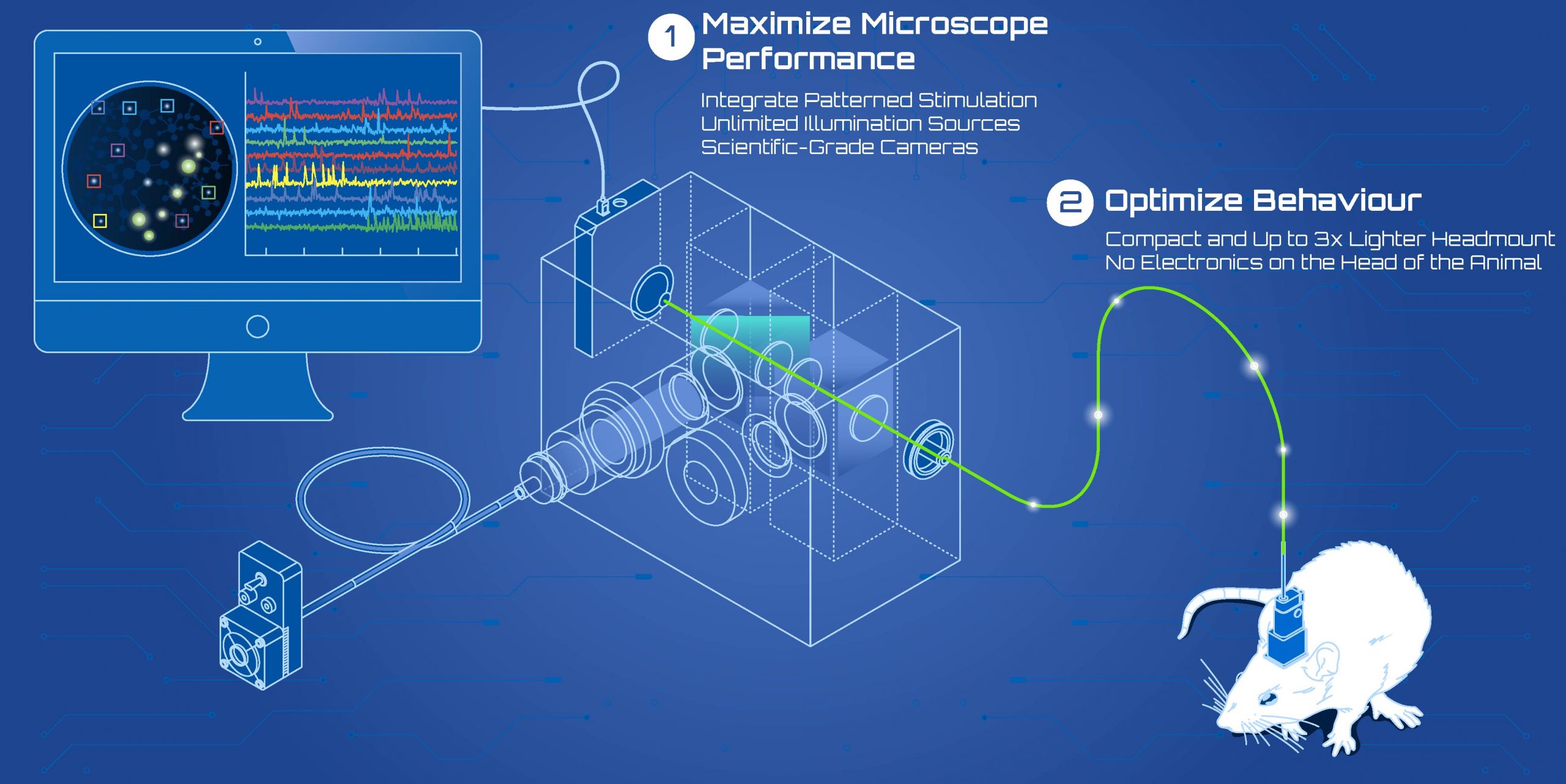
While a number of limitations have existed for optical imaging, the Canadian based company Mightex have developed a new miniscope that overcomes these hurdles. The OASIS Implant is a ground-breaking platform for simultaneous single-cell resolution optogenetics and calcium imaging in freely-behaving animals. It can image neurons in the deep-brain, cortex, or spinal cord.
Researchers can investigate a single region of the nervous system or multiple regions at the same time. The system is an all-optical solution for freely-behaving experiments in preclinical neuroscience research.
It is a reconfigurable solution that allows customizability for different research goals including different wavelegths of illumination, spatiotemporal control of stimulation, cameras, imaging fibers, and more. By adding the necessary components to the system as time progresses, the OASIS Implant can evolve as experiments evolve. With the OASIS Implant platform researchers can transition from fiber photometry functionality to cellular-resolution calcium imaging capability or from widefield optogenetic stimulation to single-cell patterned stimulation.
Are you interested in learning more about Mightex’s products? Contact us today for more information!

Green Leaf Scientific supplies a range of specialty neuroscience research products for both in vivo and in vitro studies. We have a range of suppliers such as Strex Inc., Diagnostic Biochips Inc., Pinnacle Technology Inc. and more!
Green Leaf Scientific supplies a range of advanced wireless technologies and freely-moving solutions to pre-clinical neuroscience researchers. Interested in learning about our suppliers’ products? Contact us today!